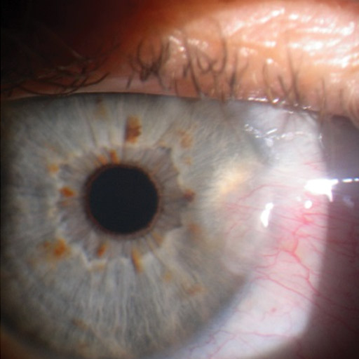
A pterygium (pronounced “te-ridge-e-um”) is an elevated wedge -shaped growth on the conjunctiva or mucous membrane that covers the white part of your eye. It usually originates from the inside corner of the eye, but less commonly from the outside corner. The word Pterygium is from the Greek word “pterygos” meaning “wing”, Typically that of a butterfly. The Ptyrigium resembles a butterfly wing in both shape and appearance. These growths are non-cancerous, but they do contain blood vessels and can form scar tissue that can permanently disfigure the eye. Exposure to ultraviolet light has been identified as the main culprit regarding the growth of a pterygium. Wind, dust, and exposure to ultraviolet light will cause the pterygium to become inflamed, dry, and very itchy. These growths need to be surgically removed by an eye specialist before they invade the cornea and affect vision. Ptyrigia that occur on the surface of the eye are commonly mistaken as cataracts which is actually inside the eye. Recurrence of the pterygium is minimised by wearing good quality sunglasses with a 100% UV filter.

Signs and Symptoms
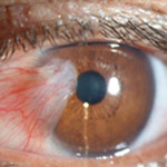
A pterygium starts as redness and thickening in the corner of the eye – usually the corner closest to the nose. The growth can extend across the surface of the eye towards the iris (the coloured part of the eye). Often a person may notice the formation of a pterygium but may not experience any other symptoms.
Typical Signs of a pterygium are:
- Wedge-shaped, translucent membrane with apex extending onto cornea.
- White to pink in color, depending on vascularity.
- Vascular straightening in the direction of the advancing head of the pterygium.
- Stocker line: iron line on cornea at leading edge of pterygium.
- Regular or irregular astigmatism
- Degenerative changes.
Symptoms may vary, but the most common are
- Eye redness and inflammation.
- A gritty feeling in the eye.
- A feeling that there is a foreign object in the eye.
- Dryness of the eye due to reduced tear production.
- Blurring of vision if the corneal surface is altered or “warped”.
- Obscuring of vision if growth encroaches across the pupil.
Causes and Risks
The cause is unclear. It appears to be partly related to long term exposure to UV light and dust. Genetic factors also appear to be involved. It is a benign growth. Other conditions that can look similar include a pinguecula, tumor, or Terrien's marginal corneal degeneration.
Risk Factors typically include:
- Ultraviolet exposure (single most significant risk factor).
- Exposure to irritants (dust, sand, wind).
- Inflammation.
- Dry ocular surface.
- A pterygium can lead to severe scarring on your cornea, but this is rare.
- Scarring on the cornea needs to be treated because it can cause vision loss.
Stats and Incidence
Pterygia occurrence is much greater among people who live near the equator. But it also can develop in anyone who lives in a sunny climate. It's most often seen in young adults aged 20 to 40. It appears to be more common in men than in women.
Pterygia are often preceded by a related non-cancerous condition called a pinguecula (pin-gwek-yoo-la). This is a yellowish patch or bump on the conjunctiva near the cornea. The conjunctiva is the thin, moist membrane on the surface of the eye.
The frequency of the condition varies from 1% to 33% in various regions of the world. It occurs more commonly among males than females and in people who live closer to the equator. The condition becomes more common with age. The condition has been described since at least 1000 BC.
In a study conducted, the pooled prevalence of pterygia was 10.2%. The pooled prevalence among men was higher than that among women (14.5% vs 13.6%). The proportion of participants with unilateral cases of a pterygium was higher than that of participants with bilateral cases of pterygia. It was found that the higher pooled prevalence of pterygia was associated with increasing geographical latitude and age in the world.
Treatment
The pterygium itself is normally harmless and treatment is purely to relieve symptoms when they occur. A pterygium often causes a dry eye problem, and most symptoms tend to be relieved by an artificial tear drop.
If the lesion causes persistent discomfort or interferes with vision, it can be surgically removed during an outpatient procedure. The operation can be performed under local anaesthesia (awake) or general anaesthesia (asleep). The choice is based on anticipated length of surgery, the size of the pterygium, the surgeon's advice and the patient's own preferences.
Surgical intervention is considered if:
- Your symptoms are not adequately relieved by the eye drops.
- Your symptoms recur frequently.
- There is pus in the eye or the eyelids stick together on waking in the morning.
- The pterygium covers part of the iris and grows onto the cornea and towards the pupil.
- You have any deterioration of vision.
- You would like the pterygium to be removed for cosmetic reasons.
The type of surgery most commonly used today uses a graft from the patient's own conjunctiva (surface eye tissue) or preserved amniotic membrane (the thin tissue forming the sac in which a foetus grows) to fill the empty space created by the removal of the pterygium. In this procedure, the pterygium is removed and the conjunctiva or amniotic membrane is glued or stitched onto the affected area.
Prevention, and specifically post operatively, wearing sunglasses and a hat if in an area with strong sunlight is essential. Following surgery, a pterygium may recur in around half of cases.
References
- Wikipedia
- Pterygium: Causes, Symptoms & Diagnosis - Healthline
- Pterygium in Indonesia: prevalence, severity and risk factors. Gazzard G1, Saw SM, Farook M, Koh D, Widjaja D, Chia SE, Hong CY, Tan DT.
- Health 24 – Eye Health
- Reviewed by Dr Clive Novis, Ophthalmologist, (November 2010)
- Cullen, A.P., Ozone depletion and solar ultraviolet radiation: ocular effects, a United Nations environment programme perspective. (2011).
- Boots WebMD
- BMJ Journals
- Southern Cross Medical Care Society
- American Academy Of Ophthalmology